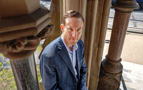
When Michael Paradiso ’81 ScM, ’84 PhD attended Pomona College in California in the 1970s, he studied physics, eventually earning a PhD at Brown in the subject. He intended to focus on high-energy subatomic particles, which even today remain the foundation for our understanding of the universe. Scientists who study them ponder such basic questions as: How did the universe come into being? What’s it made of? How long will it last?
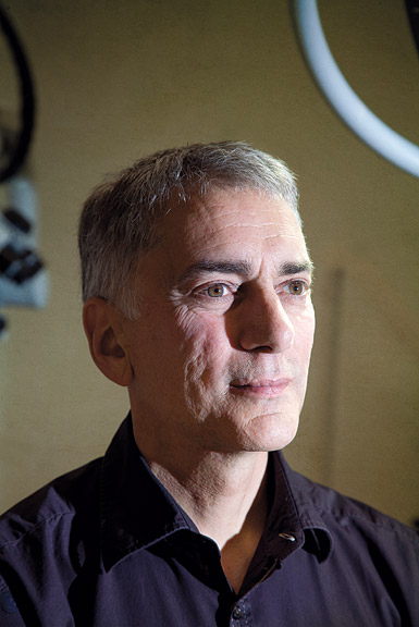
Thirty years later, Cooper had turned his attention to a new discipline: neuroscience. If there is an undertaking as intriguing and ambitious as solving the mysteries of the cosmos, he thought, it’s understanding the workings of the brain. Paradiso found Cooper’s research fascinating. He sat around at lunch with Cooper’s students discussing higher and lower brain processes and the various networks of neurons that decipher the information gathered by our senses. The questions now were: How do humans make sense of their environment? How does experience shape the development of our brains? The answers promised to explain how we become who we are—the origin of what makes us unique individuals.
These were not new issues. Philosophers and scientists had been asking them for centuries, but Paradiso believed they could be better answered by science. A whole new set of technological tools was coming online, offering the most detailed and precise images of the brain ever seen. Leading the way in the 1990s was functional magnetic resonance imaging (fMRI), which let researchers trace blood flow through the brain. Ask someone in an MRI machine to smile, to recall a bad memory, to fall asleep, or to solve a math problem, and you could observe where the blood flowed when carrying out the activity. Tying specific areas of the brain to certain brain functions now became possible.
Paradiso soon switched to studying neuroscience. “What could be more important than figuring out how the brain works?” he thought. “We know so little, the potential for discovery is vast.”
Since 2007, Paradiso has been director of Brown’s Center for Vision Research, an interdisciplinary grouping of more than forty scholars from ten different academic departments, each researching some aspect of vision. The name is really a double entendre, referring as it does to the literal sense of vision as well as the more basic question of how the human consciousness “sees,” or perceives, the world. Working at the center, which is rooted in Brown’s multidisciplinary research culture, are not only ophthalmologists looking at diseases of the eye, but philosophers and historians studying past and present theories of perception, and computer scientists trying to get machines to see like humans. And then there are the neuroscientists dedicated to understanding, neuron by neuron, what happens in the human brain when we look at the world around us.
Paradiso is surprisingly affable and easygoing for a high-profile scientist. He kicks his legs up on his desk, revealing polka-dot socks. He chose to focus on vision, he says, because it is a fundamental activity of the brain, integral to human consciousness and critical to our daily lives. About 30 to 40 percent of the brain’s 100 billion cells, or neurons, play a role in it. Vision, he explains, is more than the sensory act of seeing; it also includes the process of making meaning out of what we’ve seen. Such things as color, texture, shape, depth, context, and motion are all processed by the brain’s vision system, as is the recognition of faces and objects and the ability to distinguish foreground from background.
Vision is the main way we construct our reality, Paradiso explains, and its input drives much of how we think and feel about the world around us. What we see forms the basis of our memory, our behavior, and our awareness of ourselves in place, time, and context. For someone like Paradiso, vision is the perfect way in to studying memory and consciousness. “Vision is at the core of who we are,” he says. “I love all my senses—hearing, tasting, feeling—but vision is the sense I would least want to lose.”
The early twenty-first century is a good time to be a brain researcher. The field is now getting the kind of attention and national push that mapping the human genome got in the last few decades. This spring President Obama announced a $100 million effort to map the entire human brain, a task on which the researchers at the Center for Vision Research have long been hard at work. Using new technologies, such as microelectrodes, they can now look at brain activity with a level of detail that was not possible with MRI technology, which offered a view of the brain comparable to looking down from an airplane at a river. Using MRI, scientists could get a sense of flow and landscape, but they couldn’t clearly see individual trees, fences between houses, or stop signs.
Now, scientists can observe individual neurons working together in networks to carry out various brain functions. This means not only understanding the function of a single brain cell, but also figuring out how clusters of neurons interact to produce amazingly complex calculations and activities. It’s like being able to look at brain activity on a small and large scale at the same time.
Mapping the visual system and understanding how its cells work together would, for Paradiso and his colleagues, be an achievement comparable to mapping a portion of the human genome. They would be able to identify the neurons and neuronal pathways that enable us to perceive the world and visually construct our sense of reality. At a practical level, this knowledge would open up the possibility of developing treatments for a raft of diseases in which the visual system has been implicated: among them blindness, dyslexia, autism, schizophrenia, and Alzheimer’s. It could also lead to treatments that undo damage to the visual system caused by a stroke or brain injuries.
Answers to the big, philosophical questions that so interest Paradiso
would also start to emerge. We would at last understand what’s involved
when we think, perceive, and interpret—nothing less than what it means
to be a conscious human being.
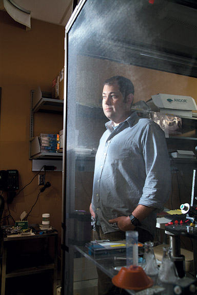
As it happens, in the first few weeks of a tadpole’s life, its vision evolves in much the same way as it does in a human fetus. Aizenman describes tadpoles as “swimming embryos,” making them perfect subjects for understanding the simplest neurological processes and how they can go awry to produce some serious diseases.
To test tadpole eyesight, Aizenman puts the animals in a standard
fish tank with a clear bottom. A computer screen beneath it displays a
series of moving black dots. The tadpoles see the dots as shadows, as
predators—as danger. A camera poised above the tank captures a single
tadpole’s reaction to the dots: when it swims away, what direction it
takes, and how far and for how long it travels. All these measurements
indicate how well the tadpole sees. The better its sight, for example,
the sooner it will spot the dot and move away.
What Aizenman does next is play games with the tadpole’s brain. Genes
provide a simple and rough blueprint for vision, whether in tadpoles or
humans. The animal’s environment then acts upon those genes to shape
the vision system. Sometimes he shines a very bright light or a
flashing strobe at the tank. Other times, there’s complete darkness.
These changes induce abnormalities. Certain connections between neurons
don’t occur. Critical patterns of neuron firings that let the tadpole
gauge depth, contrast, and shape never happen. In the tank, Aizenman
can see what’s gone wrong with their sight by observing the change in
their reactions to the black dots. They may respond later, or not at
all, suggesting something has gone haywire with their ability to gauge
distance.
After putting the tadpoles through various tests, Aizenman anaesthetizes them and examines their brains, carefully inserting a pipette into their heads that’s small enough to pierce a single one of their neurons. A sensor inside the pipette then takes measurements of the electrical current emitted by the brain cell. “We can see how one small manipulation leads to all sorts of defects,” Aizenman says. “They cascade into an abnormally wired brain.”
By correlating these deficiencies with the tadpoles’ irregular behavior in the tank, Aizenman can figure out what neurons do which functions in the brain. He identifies the precise neurons and pathways among the neurons producing a particular visual ability—acuity, for example, or the discernment of brightness, or depth perception. It all comes together to produce a map of the tadpole’s brain with unprecedented precision: more Google Street View than Google Earth.
The human implications of this work are many. Some human diseases arise from abnormalities in brain development in the womb. With autism, for example, synapses become overactive. The autistic brain makes short-range connections between cells, but has difficulty with the longer ones needed to engage in social behavior and process language. Similarly, schizophrenia likely results from faulty wiring between neurons where certain genes fail to express themselves. Several years ago, Aizenman found that putrescine, a compound that plays a role in the tadpole’s brain cell division and controlling the activity of neurons (it also causes bad breath in humans), also regulates epileptic seizures.
Thanks to these kinds of experiments, Aizenman and his colleagues can
begin to understand what goes wrong in humans afflicted by such
disorders. Their work holds out the promise of our being able to
pinpoint the moment and place in the brain where the circuitry starts
to go awry and then being able to fix it so an illness never develops.

To do this, she works with rats, whose brains contain similar components and layout for processing memory as those in human brains. In both species, the hippocampus has long been known to be the hub of memory activity. But in recent years scientists have found that adjacent structures, called parahippocampal cortices, also play an important role in memory. Burwell focuses on these cortices. She follows rats’ memories by using their vision system to test their recognition of what they’ve seen in the past.
Burwell, who speaks with a Southern twang left over from her childhood in Jackson, Mississippi, is somewhat of a late-blooming academic, having been a computer programmer for ten years before earning her PhD in biological psychology at the University of North Carolina at Chapel Hill. She decided in grad school to study memory. “It just seemed like memory makes us who we are,” she says.
Several years ago, Burwell designed a new kind of rat maze. Instead of having the animals run through a labyrinth, she created a forty-six by fifty-four inch table whose bottom is a rear projection screen. Burwell projects all sorts of images onto the maze’s floor, usually such shapes as circles, squares, triangles, and diamonds.
In one experiment, Burwell projected two black squares, one enclosing a white circle and the other a cross. A partition separated the shapes. Researchers encouraged a rat to “remember” one of the two shapes by rewarding it with a food pellet or a sip of chocolate milk whenever the animal headed toward the preferred shape. With repetition, the animal started to associate a particular shape with pleasure and made a beeline for that shape as soon as it was placed in the maze.
What happens when the rat is given several days rest and then put back in the maze? Does it retain in its memory the association between the shape and the reward? Does the association stay intact if the images are two different shapes? What if they are shaded differently or their orientation is changed? The answers reveal not just the duration and strength of the associations the rodents make, but also which characteristics of the environment the rat brain retains and which it doesn’t.
The next step is for Burwell to use tiny electrodes implanted in the rat’s brain to record the firings of neurons as the animal figures out what it is looking at and calculates where it will be heading. When the rat distinguishes between a black and a white circle, Burwell can see which cells are involved in the process of remembering the object’s color. Collected at the level of the individual neuron, this information allows Burwell to map the brain pathways associated with the remembering and identification of objects.
Burwell has recently begun a new set of experiments working with a technology called optogenetics. Using a laser attached to a tiny optical fiber able to direct light to neurons in the rat’s brain, the technique sensitizes the neurons, allowing them to be turned on or off. Burwell is interested in the neurons that tell a rat whether it sees a new or familiar object. You can tell when a rat finds something new because it will spend more time exploring it. So Burwell familiarized the rat with a specific shape. After this she used the light from the laser to trigger the neurons that activate when the rat sees a new object. She found that she could make the rat regard the shape as entirely new and unremembered.
In this way, Burwell altered a specific memory embedded in the rat’s brain. In further work, she plans to answer the next logical question: do the changes stick? Has she permanently altered the animal’s memory? If she has, she will have changed the rat’s mind for good.
Burwell focuses on basic scientific research in rats, but her work
could one day have important ethical implications for humans. Someone
who’s been through a traumatic event during childhood or a war might
want all memory of it erased. But what about erasing less traumatic
memories? What about people who still suffer from the effects of an
unhappy childhood? Could they erase memories of their parents’ divorce?
It’s still far away, but brain science may one day give us the ability
to perform “cosmetic surgery” on our minds, in essence reengineering
who we are.
Neuroscience professor David Sheinberg ’94 PhD works with monkeys. He’s
mapping their vision systems so he can do to primates what Burwell did
to the rats: change their perception of reality. But he’s not altering
memory. He wants to be able to implant an idea in a monkey’s mind that
wasn’t there before. He thinks it will help people who are blind.
In one of the warren of rooms inside his lab, a monkey sits in a chair, its eyes focused straight ahead on a computer screen. A small cross and a small T appear on the screen. When the monkey looks at the T, the reward is a sip of apple juice. The monkey looks at the T every time.
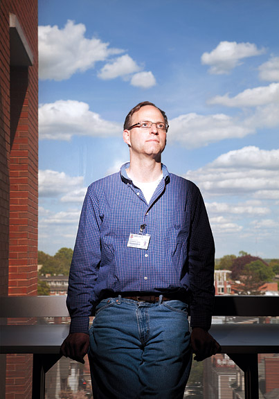
Sheinberg knows enough about the monkey’s brain that he can control where the animal directs his gaze simply by activating certain brain cells. He can now get the animal to look to the lower right of the computer screen even when the T doesn’t appear there. He is bypassing the animal’s senses and implanting an idea in his head. The monkey responds as if his brain is telling him, “Oh, there is something interesting over there. Let me look.” It doesn’t matter what his eyes see. His brain is in control.
Sheinberg says that most blind people have functioning vision systems. The piece that doesn’t work is the sensory input organ: their eyes. Sheinberg believes that if the input can enter a blind person’s brain another way, at the higher, nonsensory levels of the vision system, they might learn to experience something they’ve never actually seen. They could imagine in their “mind’s eye” the color, shape, and speed of an object, giving them, Sheinberg says, “an enhanced sense of reality.” He calls this, “knowing without seeing.”
“You may not see an object,” he says, “but you may have a sense of,
‘Oh, there’s a dog in the room,’ or ‘There’s a car over there.’”
In his office, amid the textbooks and journals, Sheinberg keeps a stack
of Daredevil comics that relate the adventures of Matt Murdock, who
runs to the rescue of a blind man about to be struck by a truck.
Radioactive material spills from the truck, blinding Murdock while
strengthening all his other senses. He can no longer see the world, but
can still imagine what it looks like. Murdock surprises his enemies by
how well he gets around.
Sheinberg doesn’t think he’ll turn anyone into a superhero. But he also doesn’t see why science can’t one day augment the vision system to give us more limited Daredevil superpowers. A golden age of brain research may soon be upon us. The potential to change who we are, how we think, and what we see is thrilling and terrifying. It is likely coming, whether we want it or not.
A Skeptical View
While the stunning advances now under way in neuroscience have unleashed a wave of excitement—and funding—from scientists, government agencies, and private industry, some observers urge caution. Among them is psychiatrist and American Enterprise Institute scholar Sally Satel ’84 MD, whose new book, Brainwashed: The Seductive Appeal of Mindless Neuro-science, cowritten with Emory psychology professor Scott O. Lilienfeld, traces neuro-science claims that the pair believes are overhyped and not supported by sound science. Satel and Lilienfeld write that, while functional MRI brain scans can tell you which parts of your brain are working harder than others, they can’t explain thoughts, feelings, the roots of addiction, or even the deeper reasons behind your preference for a particular brand of soda. While crediting neuroscientists for discoveries that can aid with certain diseases and disabilities, the authors suggest that neuroscience is only one way—not the way—of understanding how the mind works and how our perceptions of reality make us who we are.
Lawrence Goodman is the BAM’s senior writer.


